How Does the Social Security Administration Determine if I Qualify for Disability Benefits for a Stroke?
If you have had a stroke, Social Security disability benefits may be available. To determine whether you are disabled by your stroke, the Social Security Administration first considers whether your stroke and its effects are severe enough to meet or equal a listing at Step 3 of the Sequential Evaluation Process. See Winning Social Security Disability Benefits for Stroke by Meeting a Listing. If your stroke is not severe enough to equal or meet a listing, the Social Security Administration must assess your residual functional capacity (RFC) (the work you can still do, despite your stroke), to determine whether you qualify for disability benefits at Step 4 and Step 5 of the Sequential Evaluation Process. See Residual Functional Capacity Assessment for Stroke. If you have further questions about social security disability law, you may consider consulting a disability attorney. An experienced disability attorney can also help you fight back against unfair disability insurance benefit denials from major disability insurance companies.
About Stroke and Disability
What Is a Stroke?
A stroke is called a cerebrovascular accident or CVA by medical professionals. It is usually caused by either:
- Blockage of an artery in the brain by a blood clot or fatty deposits, which is called a cerebral infarction;
OR
- A ruptured cerebral artery bleeding into the brain, which is called a cerebral hemorrhage.
Some strokes are caused by cerebral aneurysms.
The Social Security Administration sees large numbers of stroke cases.
Strokes Caused by Blockage of an Artery
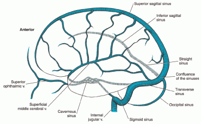
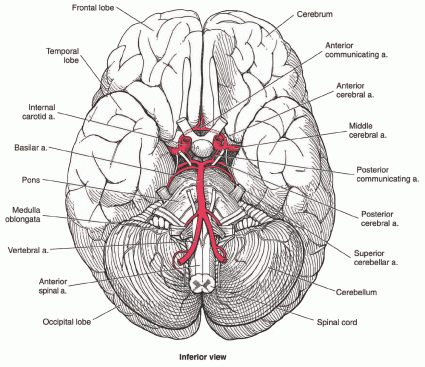
Most strokes are caused by cerebral infarction in which an artery in the brain (see Figures 1 and 2 below) is blocked depriving the brain of blood and damaging brain tissue. An arterial thrombosis (blood clot) is the most common cause of cerebral infarction. Such a clot could form in a cerebral artery itself or in the heart as a result of a variety of heart problems and be pumped to the brain. Blockage of a cerebral artery by the fatty deposits of atherosclerosis can also deprive an area of the brain of blood and lead to infarction. Actually, many cerebral infarctions are caused by cerebrovascular disease in which a blood clot forms around fatty plaques. Unlike arteries in the heart or legs, cerebral arteries cannot be cleaned out of fatty blockages. However, if a stroke occurs as a result of a blood clot, brain damage can be lessened by clot-dissolving drugs. Medical attention must be sought within a few hours for treatment to be effective and clot-dissolving drugs pose some risk of causing deadly bleeding.
A piece of atherosclerotic plaque can break off inside one of the two internal carotid arteries in the neck, be pumped to the brain, and lodge in a cerebral artery to cause an infarction.

Figure 1: Veins of the brain.

Figure 2: Base of the brain, including main arteries.
Strokes Caused by Ruptured Cerebral Artery (Cerebral Hemorrhage)
The most frequent cause of hemorrhagic CVAs is uncontrolled hypertension (high blood pressure), often related to non-compliance with medical treatment. The Social Security Administration sees many such tragic cases. Bleeding in the brain may also occur from abnormal tangles of blood vessel growths called vascular malformations and cerebral aneurysms.
Strokes Caused by Cerebral Aneurysms
Incidence of Cerebral Aneurysms
A significant number of strokes are caused by cerebral aneurysms, which are enlarged, and weak areas of a cerebral artery that can rupture and cause a subarachnoid hemorrhage (SAH). Aneurysms in the cerebral circulation are common. They are estimated to occur in between 1% and 5% of the general population and account for 5% to 15% of strokes. The most common location for cerebral aneurysms is the anterior cerebral artery. Cerebral aneurysms are twice as common in women as in men. They occur more frequently in individuals with certain disorders such as autosomal dominant polycystic kidney disease. More than one aneurysm may be present.
Millions of Americans have cerebral aneurysms. Although somewhere between 50% to 80% of aneurysms are small and do not rupture—many are only found incidentally at autopsy—that still leaves millions of individuals at risk for death or debilitating stroke.
Effects of Rupture
If a stroke occurs, the prognosis is grave, with a mortality of about 40% to 50% within 30 days of the first rupture. For surviving patients with SAH, about 30% have significant neurological abnormalities. For instance, following SAH, 15% to 20% of individuals will develop hydrocephalus (fluid accumulation on the brain) and require further neurosurgical procedures to treat that serious brain disorder.
The following scale is widely used by physicians to describe a patient’s condition after SAH:

Re-bleeding Risk
If a person has experienced one rupture (bleeding episode) from an aneurysm, the risk of future bleeding is increased to 10 times that of someone with a no rupture history. If the aneurysm was large (10 mm or more), the risk of rebleeding is even higher.
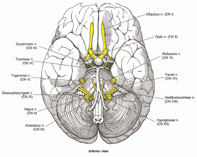
If an aneurysm has not bled previously, data indicates a low bleeding risk of 0.05% per year; in aneurysms less than 7 mm, the 5-year risk approaches zero in the absence of a bleeding history. However, the size of an aneurysm is not the only consideration—aneurysms putting pressure on vital brain structures, such as a cranial nerve (see Figure 3 below), require surgical intervention at a smaller size.

Figure 3: Cranial nerves at the base of the brain.
Diagnosis and Surgical Treatment of Cerebral Aneurysms
Many unruptured cerebral aneurysms can now be identified with CTA or MRA, without the more invasive catheter angiography. However, catheter angiography better diagnoses SAH. Angiography of any type is not perfect and can fail to identify small aneurysms of less than 3 mm.
Cerebral aneurysms may be treated surgically to reduce the risk of rupture, rebleeding, or brain damage from pressure the aneurysm places on brain tissue. Surgery to place a metal clip on the neck of an aneurysm that connects it to a parent vessel has been the standard treatment in the past. This procedure is major brain surgery and requires a craniotomy. A piece of the skull (skull flap) is sawed under general anesthesia and laid back for entry into the brain. This surgery has risks and the surgical risks for small aneurysms considerably exceed the risks of conservative (non-surgical) treatment.
A second surgical option has been the use of detachable coils of various sizes and shapes, which can be inserted without opening the skull. These coils are advanced by microcatheter to the aneurysm through the femoral artery in the leg, and then up through the carotid artery in the neck into the cerebral circulation. The coil is then detached inside the aneurysm to block blood flow through the neck of the aneurysm into its main body. Thus, the patient is spared the very invasive craniotomy. Although the risk of rebleeding after coiling is slightly greater than after clipping, the safety of coiling appears greater in many instances. Medical judgment in individual cases is still required to determine the best treatment option, but it is likely that coiling will continue to replace a significant number of cases that would otherwise have required clipping.
Diagnosis of Stroke
Evidence that a CVA has occurred is based on history and physical examination, as well as neuroimaging with computerized tomographic angiography (CTA) or magnetic resonance angiography (MRA) of the brain. Cerebral catheter angiography, a much older procedure than CTA or MRA, is still sometimes used. It carries some risk and is not needed to evaluate most CVAs. Cerebral catheter angiography involves direct injection of x-ray contrast material to outline the arteries of the brain. A catheter is threaded through the femoral artery in the leg, up into a carotid artery in the neck, and then manipulated into the cerebral circulation where contrast injection takes place.
Recovery from Stroke
Brain cells that are killed by a stroke are not replaced with new cells. The brain cannot re-grow any part of itself. But it can re-arrange brain cell connections to some degree to compensate for injury. The ability of the brain to compensate for injury decreases with age. Recovery from stroke depends on the ability of remaining brain areas to perform necessary functions, and recovery of areas not permanently damaged by the CVA. Rehabilitation is very important in achieving maximum possible recovery, and should be instituted as soon as possible after the CVA.
Effects of Stoke
CVAs can be of all degrees of severity, and the type of damage they do depends on where in the brain they occur. Some CVAs cause death immediately, while others may cause little limitation. There might be good recovery, or very little.
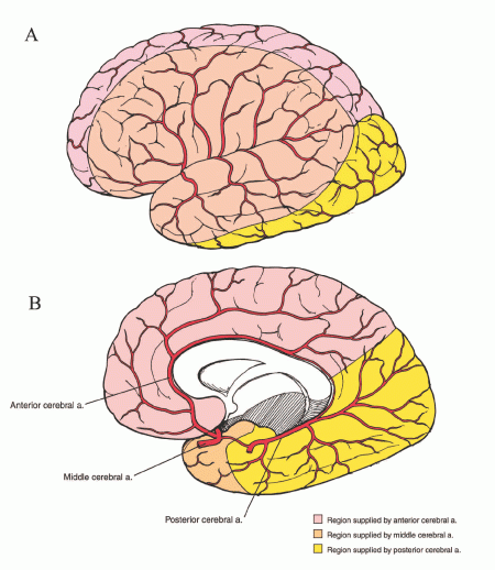
Strokes can have many effects depending on what areas of the brain are damaged (see Figure 4 below):
- Weakness, paralysis, numbness. Most CVA claimants are awarded disability benefits because of limitations in movement or motor ability, such as weakness and paralysis in an arm and leg on the same side of the body as a result of blockage (occlusion) in the middle cerebral artery or one of its branches.
- Speech and language problems. Strokes sometimes produce some degree of loss of ability to understand or express certain aspects of written or spoken language in various combinations (known as aphasia).
- Personality changes. A CVA far forward (anterior) in a frontal lobe might produce personality changes if it is large enough, without any physical limitations.
- Vision problems. A CVA might involve the occipital lobes in the back of the brain. The occipital lobes process primary visual information and a stroke in that area would produce visual losses either in acuity (sharpness) or visual fields (how wide an area a person can see) without any other impairment. Major strokes that are not in the occipital lobes may result in visual field losses in the form of loss of half of the person’s visual field. Each half of the brain carries half the total visual information. Most CVAs only involve one side of the brain and therefore at the worst can only eliminate half of a person’s visual field in a pattern called homonymous hemianopsia0.
- Balance problems. Strokes affecting the parietal lobes of the brain can produce distortions in the mental construction of space and cause loss of an awareness of body parts, a condition known as unilateral neglect. It is important that the neurological examination of a patient after a CVA detect unilateral neglect, because non-awareness of a limb makes it as functionally useless as paralysis. Strokes affecting the posterior circulation to the cerebellum can affect balance and ability to walk without producing any actual weakness.

Figure 4: The Circle of Willis, showing main cerebral arteries and the parts of the brain they supply.
Winning Social Security Disability Benefits for Stroke by Meeting a Listing
To determine whether you are disabled at Step 3 of the Sequential Evaluation Process, the Social Security Administration will consider whether your stroke is severe enough to meet or equal the stroke listing. The Social Security Administration has developed rules called Listing of Impairments for most common impairments. The listing for a particular impairment describes a degree of severity that Social Security Administration presumes would prevent a person from performing substantial work. If your stroke is severe enough to meet or equal the stroke listing, you will be considered disabled.
If you believe you are disabled because of a stroke, the Social Security Administration will not evaluate your case until at least 3 months have passed since the stroke occurred. It takes at least this long to find out how much you will recover. Exceptions could be claimants with such extreme brain damage proven by physical examination, or brain scans that they are comatose and not likely to survive or show any significant recovery in 3 months. It would be pointless to actually hold such cases for 3 months. However, in most cases prediction of the degree of recovery from a CVA is unreliable.
If your stroke caused or worsened a mental impairment, your mental impairment will evaluated under the mental impairment listings. The most likely emotional problems post-CVA are depression and organic brain syndrome. Mental impairments post-CVA is an area that the Social Security Administration is particularly likely to overlook.
The listing for stroke is 11.04, which has two parts: A and B. Most post-CVA claimants who meet the listing do so under part B, rather than part A.
Meeting Social Security Administration Listing 11.04A for Stroke
You will be disabled under Listing 11.04A if you have had a stroke(central nervous system vascular accident) with sensory or motor aphasia resulting in ineffective speech or communication more than 3 months after your stroke.
What Is Aphasia?
Aphasia means difficulty in comprehension or expression of language.
Types of Aphasia
There are many types of aphasia.
- In sensory aphasia, a person may be unable to understand written or spoken words.
- In motor (expressive) aphasia, the person knows what he wishes to say, but is unable to form and speak the necessary words, or only with extreme difficulty and slowness. Or the motor aphasia might involve inability to put thoughts into writing.
- In global aphasia, receptive and expressive language ability is essentially non-existent. In other words, the person can neither understand words, nor speak.
- Broca’s (non-fluent) aphasia is characterized by a difficulty in the production of speech, which is accomplished by only several words at a time in a halting manner; writing is also impaired, but the person may be able to understand speech and to read.
- In mixed, non-fluent aphasia, the person has difficulty with speech, understanding, writing, and reading.
- In anomic aphasia, the person has difficulty finding the right nouns or verbs to use in speaking or writing.
- In Wernicke’s aphasia, a type of fluent aphasia, the person has little difficulty in producing speech in a mechanical sense, but does not understand the meaning of words, which results in inappropriate speech, and the claimant’s ability to read and write is also severely impaired.
How Severe Must Aphasia Be to Meet Part A
All degrees of sensory and motor aphasia are possible, but to fulfill part A, the aphasia must be so severe that communication with other people is ineffective. This means that useful verbal communication is not possible because there is severe impairment in the clarity, content, or sustainability of speech. Since 90% of people have their language processing abilities in the left side of the brain, a left brain stroke is more likely to produce problems with aphasia. Although many people with CVAs have some degree of aphasia, few have it so severely that they qualify under part A.
If the aphasia is so severe that the claimant is incapable of any communication, part A is obviously satisfied. But aphasia is usually only partial. In these instances, careful examination by a neurologist or psychiatrist can provide useful clinical information regarding severity. A formal evaluation of speech communication ability can be obtained from speech therapists.
Meeting Social Security Administration Listing 11.04B for Stroke
You will be disabled under this listing if you have had a stroke (central nervous system vascular accident) and more than 3 months afterwards you have significant and persistent disorganization of motor function in two extremities, resulting in sustained disturbance of gross and dexterous movements, or gait and station. Although nearly all CVAs seen by the Social Security Administration involve the brain, it is possible for there to be a CVA involving an artery supplying the spinal cord, which is also a part of the central nervous system. This event is most likely with spinal cord trauma or tumors. Since there is no brain involvement, spinal cord strokes are evaluated under part B.
Two Extremities Must Be Impaired
Part B identifies the two major areas of motor limitations as:
- Upper extremity (arm and hand) function including:
- Gross movements.
- Dexterous movements.
- Lower extremity (leg) function including:
- Gait – the manner of walking.
- Station – the manner of standing, i.e., ability to maintain a posture.
Two extremities must be involved to satisfy part B: both legs, both arms, or one arm and one leg.
Almost all stroke cases affect an upper and lower extremity on the same side of the body. When one side of the brain is damaged, any abnormalities like paralysis are going to be on the opposite side of the brain affected by the stroke. This is because the left side of the brain controls the right side of the body and vice versa. A stroke in the left hemisphere of the brain will produce deficits in the right side of the body and vice versa. A stroke on the side of the brain opposite from the dominant hand will result in greater loss of function than a stroke affecting the non-dominant hand. For example, if you are right handed, a stroke on the left side of the brain affecting use of your right hand will be more disabling than a stroke on the right side of the brain affecting your left hand.
If a lower extremity alone (i.e., a single leg) is affected, the listing cannot be met. However, if the lower extremity is so weak that a hand-held assistive device such as a quad cane is necessary for walking, then use of an arm would be tied up, with the same functional result as paralysis of the arm. Such cases would equal the listing.
If you cannot stand and walk 6 to 8 hours daily and have any significant weakness or inability to manipulate objects with an upper extremity, part B is satisfied. Inability to stand or walk for such prolonged times does not necessarily depend only on lower extremity strength. An uncoordinated gait, or poor balance that results in an unreasonably slow pace is still not a functional gait from a work perspective and would meet the listing. This is particularly important if you have neurological dysfunction in both lower extremities, but not in the arms.
Evaluating Strength
At a physical examination, a doctor can obtain information regarding strength in the upper and lower extremities with some simple and routine maneuvers. First, the examining physician can estimate strength loss by comparing the strength of muscle contraction on your weak side to your normal side. Physicians frequently report subjective determination of strength by using a scale of 0 to 5 with 5 being normal. For example a doctor might ask you to try to straighten out your leg while the doctor resists the movement. A weaker leg might be reported as 3/5 for the quadriceps muscle (muscle on the front of the thigh) compared to an expected normal of 5/5. Zero means no movement. One means a trace of movement. Two means movement with the help of gravity. Three means movement is possible against gravity, but not against resistance. Four means movement against gravity and resistance by the examining physician. Five means normal. Errors in this type of subjective testing can be caused by the person’s motivation, variations among people in “normal” strength, and differences in the doctor’s subjective assessment.
More objectively, a doctor can test your ability to walk on your heels and toes and squat and arise. Ability to walk on the toes means you can lift your body weight by contraction of your gastrocnemius (calf) muscles and implies significant strength. Ability to walk on the heels indicates that the muscles in the anterior (front) leg (opposite the calf muscles) that flex the foot still have good strength. Ability to squat and arise implies good strength of the quadriceps muscles of the thighs.
Determination of lower extremity strength after a stroke is very important, because if a person cannot stand 6 to 8 hours a day and an upper extremity is significantly affected then the listing will be fulfilled. If you cannot stand or walk 6 to 8 hours a day, you can do only sedentary work, but if you also have limited use of an upper extremity, you cannot even do sedentary work. If you cannot walk on heels or toes in the affected lower extremity after a CVA, it is not reasonable to expect that you would have the strength to stand 6 to 8 hours daily. If you can do such testing well, it is not likely you will qualify under listing 11.04B on the basis of strength deficit. However, you still may qualify based on severe lack of balance or coordination in walking.
Evaluating Walking and Balance
In evaluating how well you stand and walk, the Social Security Administration requires a detailed description from a medical doctor. This involves factors such as how weak your legs are, how much difficulty you have in keeping your balance, how fast you walk, how much help you need walking, and so forth. To meet the listing, you do not have to use a cane, crutch, brace, walker, or other assistive device to move about. Of course, if you need such devices, that would indicate a very severe limitation. However, the use of an assistive device such as a cane or crutch, or even a wheelchair, does not establish a severe limitation without supporting objective medical data. This is true, even if your treating doctor says you need the device.
After a severe CVA, walking (gait) and standing (station) may be affected in characteristic ways. If a leg and arm are weak from a CVA, a person will often walk with a circumducting gait. The weak leg moves forward with a circular motion and the weak arms has a short swing. On the other hand, a CVA in the brainstem or cerebellum may produce an unsteady (ataxic) gait, frequently with legs wide-based in an effort to compensate for the loss of balance. Your balance can be tested in neurological examination not only by observing your normal attempts at walking, but also by testing your ability to walk heel to toe. If there is weakness of the pelvic musculature, a waddling gait may result. A foot drop tends to cause a steppage gait, lifting the foot high to clear the toes from the ground. Spasticity, produced by increased muscle tone, can produce a stiff limb with inflexible movements.
Evaluating Arm and Hand Use
The use of the arms and hands is evaluated by looking at gross and dexterous movements. Dexterous movements are those like writing, picking up small objects, or other types of fine movements. Gross movements involve larger motions with the arms and hands. Movements can be influenced by weakness, or loss of control over the way an arm or hand moves, such as tremors or poor coordination. The ability to oppose fingertips to the thumb successively can give some idea as to the intactness of fine manipulatory (dexterous) ability. However, if you can do finger-thumb opposition only slowly and clumsily, then you should not be considered capable of dexterous movements. Also, your activities of daily living (ADLs) can be helpful in assessing functional severity when they describe specific inabilities or abilities (e.g., turning doorknobs, dressing, climbing stairs, shaving, etc.).
Medical Documentation of Stroke and Its Effects
You cannot convince the Social Security Administration that you are disabled just because you appear to have weakness, poor coordination, or impaired gait because all of these findings are under your control. Thus, your medical history is very important. Claimants who have actually had a severe CVA have been hospitalized. By obtaining the records from that hospitalization, the Social Security Administration can obtain a much better picture of clinical severity. The initial physical examination can be reviewed, along with the degree of recovery at the time of hospital discharge. Almost certainly, a CT or MRI scan was done of the brain and sometimes cerebral angiography. These studies can be most helpful in determining the location and severity of brain damage, so that it can be correlated with the clinical findings at the time of adjudication. Some claimants allege abnormalities that were not present at the time of their CVA and do not correspond to the location or magnitude of the initial brain lesion, or they seem to manifest weakness much greater than that present in their past medical records. If the claimant underwent rehabilitation in the past, that information is useful.
Certain objective signs are present after a CVA. Deep tendon reflexes (DTRs) in the affected parts of limbs should be hyperactive, or faster than normal. For example, tapping the patellar tendon below the knee (knee-jerk reflex) would cause the quadriceps muscle to more rapidly contract and the leg to extend more quickly than normal.
Reflexes are graded from 0 to 4. Zero (0) means no reflex. One (1) means a distinctly hypoactive (slow) and abnormal reflex. Two (2) andthree (3) mean a normal reflex. Four (4) means a distinctively hyperactive and abnormal reflex. Physicians usually report reflexes in a right/left mode. For example, a knee-jerk (KJ) reflex reported as 4+/2 indicates a very pathological KJ on the right. Reports of 2/2 or 3/3 for reflexes should be interpreted as normal. However, asymmetry in reflexes may be significant, e.g., 2/3+ might indicate an abnormal reflex on the left.
Additional basic reflexes important in neurological examination include the achilles reflex (AJ), biceps jerk (BJ), triceps jerk (TJ), and brachioradialis reflex. There are others that might be important in particular circumstances. Also, there may be a positive Babinski sign, which means the great toe in the affected lower extremity moves upward when the bottom lateral surface of the foot is stroked. Normally, the great toe moves downward with this test. Therefore, when an examining doctor says there is a positive Babinski, this is an indication of some type of upper motor neuron lesion. An upper motor neuron lesion may also produce sustained clonus, which is a rhythmic relaxation and contraction of a muscle caused by suddenly stretching a tendon. Clonus is most frequently tested for by a doctor quickly pushing the foot upward to stretch the achilles tendon. Sustained beating movements of the foot are an abnormal clonus indicative of upper motor neuron disease. In addition, muscle tone is increased in upper motor neuron lesions, and, if marked, results in spasticity.
Residual Functional Capacity Assessment for Stroke
What Is RFC?
If your stroke is not severe enough to meet or equal a listing at Step 3 of the Sequential Evaluation Process, the Social Security Administration will need to determine your residual functional capacity (RFC) to decide whether you are disabled at Step 4 and Step 5 of the Sequential Evaluation Process. RFC is a claimant’s ability to perform work-related activities. In other words, it is what you can still do despite your limitations. An RFC for physical impairments is expressed in terms of whether Social Security Administration believes you can do heavy, medium, light, or sedentary work in spite of your impairments. The lower your RFC, the less the Social Security Administration believes you can do.
Limitations on Lifting and Walking
When assessing your RFC, the Social Security Administration should consider the weight that you are able to lift and carry. To be able to do medium work, which requires you to lift and carry up to 50 lbs and stand and walk 6 to 8 hours daily, you should have no more than very modest deficits in strength, coordination, and balance.
If you have some difficulty in walking on your heels and toes, or squatting and arising, during medical examination, you might still be able to do light work (lifting no more than 20 lbs and still standing and walking 6 to 8 hours daily). But you probably would not realistically be able to do even light work if you cannot walk on your heels and toes while carrying no weight during a physical.
In evaluating upper extremity function, the Social Security Administration should consider whether you have insufficient strength to operate arm or leg controls more than occasionally in the affected limbs.
Of course, all factors have to be considered: where your CVA is located, how large it is, whether your muscles appear weak and limp on exam or spastic (in spasm), whether you are overweight etc. Thus, your RFC could be reduced to sedentary work requiring no more than 2 hours standing and walking daily. If you also have significant upper extremity dysfunction, you would be disabled under a listing.
If both your legs are impaired, and your upper extremities are functionally intact, the impairment will meet Listing 11.04B and your will be disabled if your gait and station are impaired to a significant degree—that is, if you cannot stand and walk 6 to 8 hours daily, and you have a problem standing because of balance insufficient for sedentary work. If gait and station are fully functional for sedentary work, except for enough stamina or strength to stand and walk for more prolonged periods, and your upper extremity function is fully intact, then you are capable of a sedentary work RFC.
Limitations on Activities of Daily Living
You—or family members or care givers as appropriate—should be asked in detail about your ability to carry out activities of daily living. Of particular interest is your ability to walk up and down steps, the speed at which you walk, and how easily you tire. If you cannot walk a block, you certainly cannot stand and walk 6 to 8 hours daily. You should also be asked what tasks you could do before your stroke that you cannot do now. Can you dress without assistance? Manipulative functions are important. Can you turn a doorknob? Can you pick up coins and button shirts?
Cerebral Aneurysms and Vascular Malformations
There are no absolute rules for assessing the RFC of persons with cerebral aneurysms and vascular malformations. Assessing these impairments involves considerable medical judgment. Here are some guidelines that Social Security Administration ought to follow.
If you have an un-operated cerebral artery aneurysm or vascular malformation that does not meet or equal a listing, you should not be given higher than a RFC for light work even if you have no neurological abnormalities. This RFC would be given to minimize the risk of a hemorrhagic CVA that might be caused by heavier work that would raise your blood pressure. The risk is even greater if you have uncontrolled high blood pressure (hypertension), multiple or large aneurysms or vascular malformations.
If your aneurysm or vascular malformation has bled in the brain and definitive surgical repair was not possible, the RFC should not be higher than sedentary work or at most, light work, even if you have no neurological deficits. Obviously, if an intracranial bleed occurred spontaneously then it has a high risk of recurrence and exertion should be minimized. There is no scientific data regarding what level of exertion is safe for people with cerebral aneurysms and vascular malformations because no one would be crazy enough to create such a study or enroll in it.
Un-operated cerebral artery aneurysms or vascular malformations that have not bled and do not meet or equal a listing are difficult to evaluate. If the aneurysm is large—10 mm in diameter or more—you should not be given higher than an RFC for light work even if you have no neurological abnormalities. This RFC would be given to minimize the risk of a hemorrhagic CVA that might be caused if the aneurysm ruptures due to increases in blood pressure during exercise. The risk is even greater if you have uncontrolled hypertension (high blood pressure), multiple or large aneurysms or vascular malformations. Since hypertension is a risk factor for strokes, it is not unreasonable to suspect that higher blood pressures increase the risk of bleeding from cerebral aneurysms.
A person with a small aneurysm of 7 mm or less and no neurologic abnormalities and no uncontrolled hypertension might not be functionally restricted, while a 7 mm aneurysm in the presence of significant hypertension might justify a restriction to medium work. There is no way to avoid medical judgment in this area. There is no study showing that small cerebral aneurysms that have never bled produce any functional restriction or are more likely to bleed with normal blood pressure and even heavy exercise. In fact, since many such aneurysms are discovered incidentally, a significant number of people must have engaged in heavy exertion of various types without ill results.
If you have a large cerebral aneurysm—say 7 mm or more—and have had surgical clipping or coiling after an episode of bleeding (subarachnoid hemorrhage), your RFC would probably be medium work or no limitation, if you are otherwise asymptomatic without uncontrolled high blood pressure. If bleeding had occurred previously, or if you have uncontrolled hypertension, light work or medium work would be more appropriate even if surgery has been done—provided there was good surgical control of the aneurysm.
Breathing Issues
Most neurologists performing examinations on post-CVA claimants do not even comment on the possibility of paralysis of the diaphragm, although it can significantly affect ability to breath. This area is often not given sufficient consideration by Social Security Administration adjudicators. If the medical evidence is not clear, you and your doctors should be asked about any breathing difficulties you have. Pulmonary function studies can make the difference between allowance and denial.
Subtle Abnormalities
Some neurologists do not check claimants for more subtle abnormalities that could have significant impact on RFC, such as the ability to identify an object placed in the hand without looking at it. You and your family members should bring to your doctor’s attention any information you may have about subtle, but important deficits like this.
Facial Paralysis
Facial paralysis to various degrees occurs fairly frequently. Drooling, loss of facial expression, speech articulation problems, and difficulty eating are some of the functional problems that can result from a CVA that would affect RFC.
Getting Your Doctor’s Medical Opinion About What You Can Still Do
Your Doctor’s Medical Opinion Can Help You Qualify for Social Security Disability Benefits
The Social Security Administration’s job is to determine if you are disabled, a legal conclusion based on your age, education and work experience and medical evidence. Your doctor’s role is to provide the Social Security Administration with information concerning the degree of your medical impairment. Your doctor’s description of your capacity for work is called a medical source statement and the Social Security Administration’s conclusion about your work capacity is called a residual functional capacity assessment. Residual functional capacity is what you can still do despite your limitations. The Social Security Administration asks that medical source statements include a statement about what you can still do despite your impairments.
The Social Security Administration must consider your treating doctor’s opinion and, under appropriate circumstances, give it controlling weight.
The Social Security Administration evaluates the weight to be given your doctor’s opinion by considering:
- The nature and extent of the treatment relationship between you and your doctor.
- How well your doctor knows you.
- The number of times your doctor has seen you.
- Whether your doctor has obtained a detailed picture over time of your impairment.
- Your doctor’s specialization.
- The kinds and extent of examinations and testing performed by or ordered by your doctor.
- The quality of your doctor’s explanation of your impairment.
- The degree to which your doctor’s opinion is supported by relevant evidence, particularly medically acceptable clinical and laboratory diagnostic techniques.
- How consistent your doctor’s opinion is with other evidence.
When to Ask Your Doctor for an Opinion
If your application for Social Security disability benefits has been denied and you have appealed, you should get a medical source statement (your doctor’s opinion about what you can still do) from your doctor to use as evidence at the hearing.
When is the best time to request an opinion from your doctor? Many disability attorneys wait until they have reviewed the file and the hearing is scheduled before requesting an opinion from the treating doctor. This has two advantages.
- First, by waiting until your attorney has fully reviewed the file, he or she will be able to refine the theory of why you cannot work and will be better able to seek support for this theory from the treating doctor.
- Second, the report will be fresh at the time of the hearing.
But this approach also has some disadvantages.
- When there is a long time between the time your attorney first sees you and the time of the hearing, a lot of things can happen. You can improve and go back to work. Your lawyer can still seek evidence that you were disabled for a certain length of time. But then your lawyer will be asking the doctor to describe your ability to work at some time in the past, something that not all doctors are good at.
- You might change doctors, or worse yet, stop seeing doctors altogether because your medical insurance has run out. When your attorney writes to a doctor who has not seen you recently, your attorney runs the risk that the doctor will be reluctant to complete the form. Doctors seem much more willing to provide opinions about current patients than about patients whom they have not seen for a long time.
Here is an alternative. Suggest that your attorney request your doctor to complete a medical opinion form on the day you retain your attorney. This will provide a snapshot description of your residual functional capacity (RFC) early in the case. If you improve and return to work, the description of your RFC provides a basis for showing that you were disabled for a specific period. If you change doctors, your attorney can get an opinion from the new doctor, too. If you stop seeing doctors, at least your attorney has one treating doctor opinion and can present your testimony at the hearing to establish that you have not improved.
If you continue seeing the doctor but it has been a long time since the doctor’s opinion was obtained, just before the hearing your attorney can send the doctor a copy of the form completed earlier, along with a blank form and a cover letter asking the doctor to complete a new form if your condition has changed significantly. If not, your attorney can ask the doctor to send a one-line letter that says there have been no significant changes since the date the earlier form was completed.
There are times, though, that your attorney needs to consider not requesting a report early in the case.
- First, depending on the impairment, if you have not been disabled for twelve months, it is usually better that your attorney wait until the twelve-month duration requirement is met.
- Second, if you just began seeing a new doctor, it is usually best to wait until the doctor is more familiar with your condition before requesting an opinion.
- Third, if there are competing diagnoses or other diagnostic uncertainties, it is usually best that your attorney wait until the medical issues are resolved before requesting an opinion.
- Fourth, a really difficult judgment is involved if your medical history has many ups and downs, e.g., several acute phases, perhaps including hospitalizations, followed by significant improvement. Your attorney needs to request an opinion at a time when the treating doctor will have the best longitudinal perspective on your impairment.

